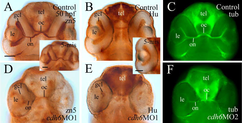
Figure 8.
Analysis of RGC differentiation using zn-5 (panels A, D and panel A insert), anti-HuC/HuD (panels B, E and panel B insert) and anti-acetylated tubulin (panels C and F) immunostaining. All images are ventral views of whole mount heads (anterior up) or eye (panel B insert). Panels A, B, D and E are processed using immunoperoxidase methods, while panels C and F are processed using immunofluorescent methods. Abbreviation: oc, optic chiasm; tel, telencephalon. Other abbreviations are the same as in Figure 1. Scale bars = 50 μm.

