
Fig. 5.
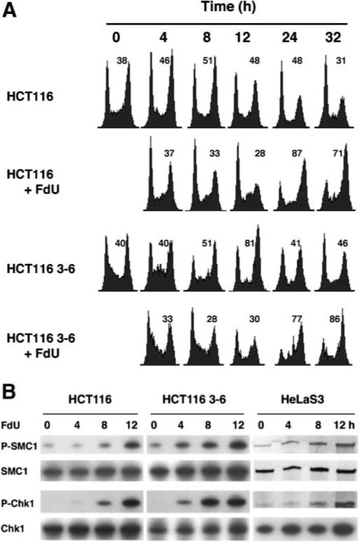
Cell cycle arrest and ATR kinase activation in response to continuous exposure to FdU. A: Kinetics of the G2/M cell cycle arrest in cells after continuous exposure to FdU. Sixteen hours after release from serum starvation, HCT116 and HCT116 3−6 cells were exposed to 0.25 μM FdU, denoted as 0 h. Cells were harvested at indicated times and cell cycle profiles were assayed by FCM. The percentage of cells in G2 is denoted in the upper right of each histogram. B: MMR-independent ATR kinase activation induced by FdU after continuous exposure to FdU. HCT116 and HCT116 3−6 as well as HeLaS3 cells were treated with 0.25 μM FdU continuously for 4, 8, or 12 h and harvested. Cell lysates were subject to Western blotting using indicated antibodies.