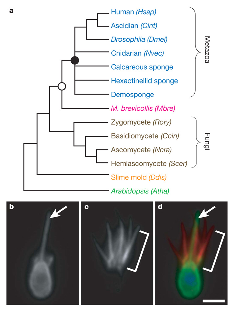
Figure 1. Introduction to the choanoflagellate Monosiga brevicollis.

a, The close phylogenetic affinity between choanoflagellates and metazoans highlights the value of the M. brevicollis genome for investigations into metazoan origins, the biology of the last common ancestor of metazoans (filled circle) and the biology of the last common ancestor of choanoflagellates and metazoans (open circle). Genomes from species shown with their abbreviation were used for protein domain comparisons in this study: human (Homo sapiens, Hsap), ascidian (Ciona intestinalis, Cint), Drosophila (Drosophila melanogaster,Dmel), cnidarian (N. vectensis, Nvec), M. brevicollis (Mbre), zygomycete (Rhizopus oryzae, Rory), basidiomycete (Coprinus cinereus, Ccin), ascomycete (Neurospora crassa, Ncra), hemiascomycete (Saccharomyces cerevisiae, Scer), slime mould (Dictyostelium discoideum, Ddis) and Arabidopsis (Arabidopsis thaliana, Atha). b–d, Choanoflagellate cells bear a single apical flagellum(arrow, b) and an apical collar of actin-filled microvilli (bracket, c). d, An overlay of β-tubulin (green), polymerized actin (red) and DNA localization (blue) reveals the position of the flagellum within the collar of microvilli. Scale bar, 2 µm.
