
Figure 3.
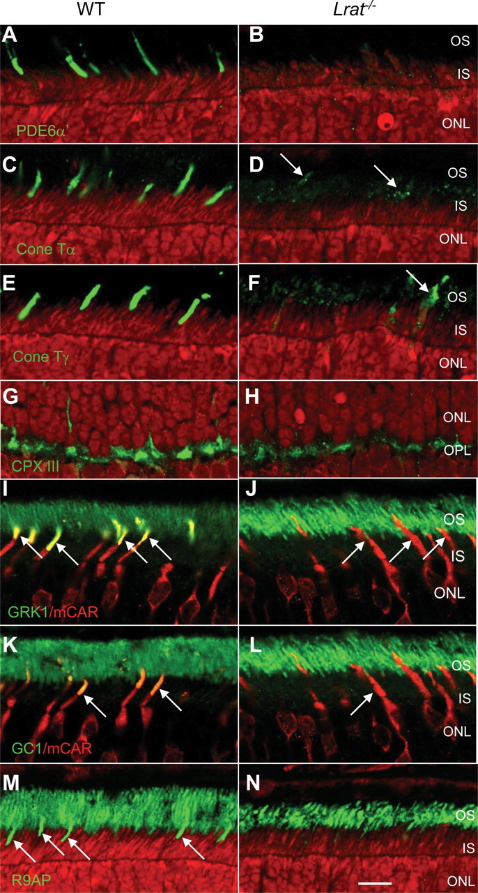
Immunolocalization of peripheral membrane-associated proteins, GC1, and R9AP in P15 WT and Lrat−/− retinas. P15 littermate WT and Lrat−/− eyes were coembedded, and central retina sections were probed simultaneously. Sections of P15 WT and Lrat−/− retinas were probed using anti-PDE6α′ (A, B), anti-cone Tα (C, D), anti-cone Tγ (E, F), anti-CPX III (G, H), anti-GRK1 (I, J), anti-GC1 (K, L), and anti-R9AP (M, N) antibodies. Bright red nuclei in B and H indicate apoptotic photoreceptors. I–L, Central retina sections were probed simultaneously with either monoclonal anti-GRK1 antibody (I, J, green) or anti-GC1 antibody (K, L, green) and polyclonal anti-cone arrestin antibody to identify cones (I–L, red). Colocalization of mCAR with GRK1 (I, yellow at arrows) or GC1 (K, arrows) is evident in WT COSs, whereas in Lrat−/− sections, COSs are largely devoid of GRK1 (J) and GC1 (L).