
Figure 3. Kinase activity of Four-jointed.
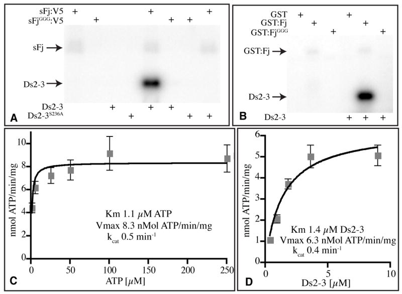
A, B) Autoradiograms of protein gels dispalying the products of in vitro kinase reactions with the indicated enzymes (top) and substrates (bottom). The positions of Ds2-3:FLAG and sFj:V5 or GST:Fj are indicated. A) Phosphorylation of Ds2-3, but not Ds2-3S236A, by Fj in vitro. B) Kinase activity of GST:Fj isolated from E. coli. C) Reaction velocity versus ATP concentration, using 2.5 μg Ds2-3:FLAG, 10 ng sFj:V5, and 1, 5, 25, 50, 100, or 250 μM ATP for 15 min. G) Reaction velocity versus Ds2-3 concentration, using 500 μM ATP, 10 ng sFj:V5, and 0.36, 0.90, 1.8, 3.6, or 9.0 μM Ds2-3:FLAG for 15 min. Plots in C and D show average values from four experiments, error bars indicate s.e.m.