Fig. 4.


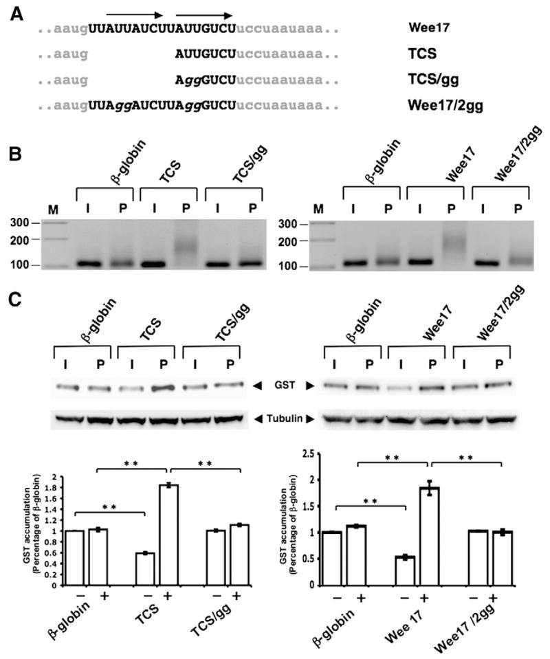
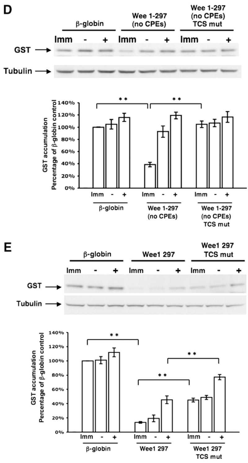
Disruption of translational control sequence (TCS) function within the Wee17 region. (A) Schematic representation of the nucleotide disruptions used to inactivate the two TCS elements (arrows) within the Wee17 region. (B) Progesterone-dependent polyadenylation of wild-type or mutant TCS reporter mRNAs. Experiments were performed essentially as described in the legend to Fig. 3, using a forward primer specific to the β-globin 3′ UTR of the injected reporter mRNAs. Oocytes were cultured in the presence of progesterone for 5 h. (C) Translational regulation of the wild-type or mutant TCS-containing β-globin 3′ UTR GST reporter mRNAs was assessed by western blotting (the β-globin 3′ UTRs analyzed had either a single TCS element (left panels) or two TCS elements (right panels)). The same lysates were analyzed for tubulin protein levels as an internal control for protein loading. The levels of GST accumulation were quantitated over three independent experiments (graph). Error bars represent SEM. One-way analysis of variance and post hoc Newman–Keuls multiple comparison test were performed. Differences between the means were scored as statistically significant, P<0.001 (**). Oocytes were cultured in the presence of progesterone for 5 h 30 min (left panel) or 5 h (right panel). (D) Translational regulation of GST reporters fused to either the entire Wee1 3′ UTR that lacked CPE sequences (Wee 1–297 (no CPEs); see Fig. 1) or the Wee 1–297 (no CPE) 3′ UTR that also had mutations in both TCS elements (Wee 1–297 (no CPEs) TCS mut) was assessed by western blotting. The same lysates were analyzed for tubulin protein levels as an internal control for protein loading. The levels of GST accumulation were quantitated over three independent experiments (graph) as described for panel C. Oocytes were cultured in progesterone for 5 h (when they reached GVBD50) and segregated into those oocytes that had (+) or had not (−) completed GVBD. (E) Translational regulation of GST reporters fused to either the entire wild type Wee1 3′ UTR (Wee1 297, see Fig. 1) or the Wee1 297 3′ UTR that also had mutations in the two TCS elements (Wee1 297 TCS mut) was assessed by western blotting. The same lysates were analyzed for tubulin protein levels as an internal control for protein loading. The levels of GST accumulation were quantitated over three independent experiments (graph) as described for panel C. Oocytes were cultured in progesterone for 5 h (when they reached GVBD50) and segregated into those oocytes that had (+) or had not (−) completed GVBD.
