
Fig. 1.
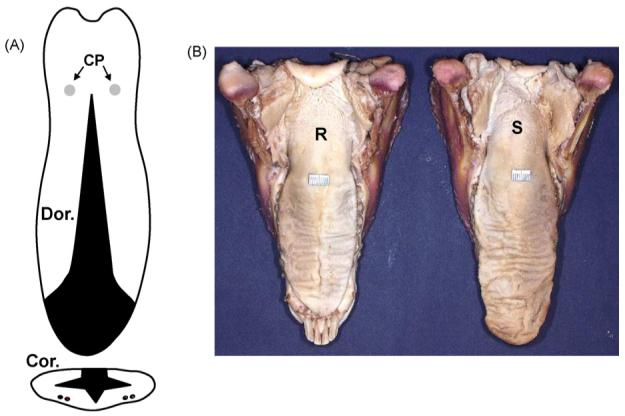
(A) Schema of the tongue reduction surgery on dorsal (Dor.) and coronal (Cor.) views. Black areas indicate removed tongue tissue. Two dots in each side indicate the locations of neurovascular bundles in the ventral surface. CP: circumvallate papillae. (B) Postmortem tongue specimens 4 weeks after surgery. R: reduction tongue; S: sham tongue.