
Figure 1.
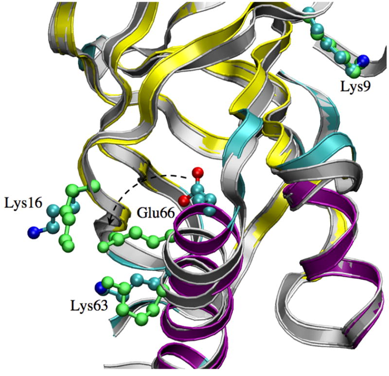
Structural features of the V66E SNase during pKa simulations (Set A-V66E). A snapshot in the λ = 1.0 window (in grey ribbon) is overlapped the X-ray structure (ribbon, color coded by secondary structure: purple: helix; cyan: turns; yellow: β-sheet). The sidechains of Glu66 and a number of important charged groups are shown in the CPK form; those colored by atom type are in the X-ray structure, and the green ones are for the λ = 1.0 snapshot. Note that Glu66 undergoes a rotation towards the protein surface.