Figure 9.

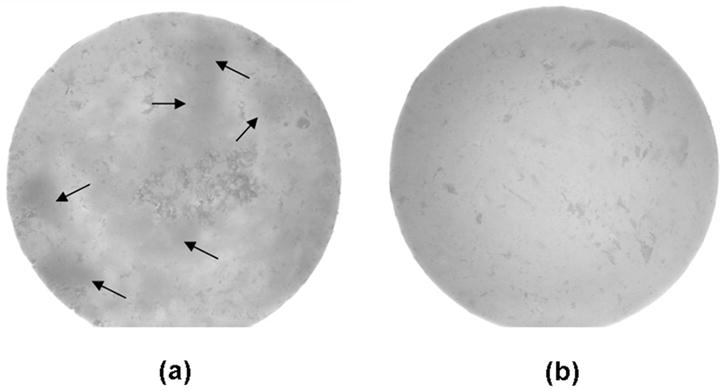
Pictures of unmilled amorphous calcium phosphate (ACP) (A) and milled ACP (B) composite disks taken using a stereomicroscope. Arrows indicate examples of filler-rich areas within the composite.

Pictures of unmilled amorphous calcium phosphate (ACP) (A) and milled ACP (B) composite disks taken using a stereomicroscope. Arrows indicate examples of filler-rich areas within the composite.