
Figure 1. Mammary tumors in MMTV-c-Neu mice have activated mTOR signaling.
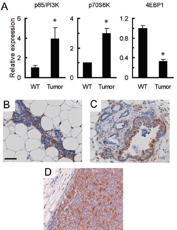
A. Gene expression microarray analysis indicates that mRNA levels for components of the PI3-Kinase and mTOR signaling pathways are altered in MMTV-c-Neu tumors. As previously described, age-matched wild type glands (WT, n=3 pools of 5 mice each) were collected and compared to primary tumors (n=7) (19). Mean signal intensities from microarrays were normalized to wild-type levels, and the normalized values are displayed on the y-axis with tissue type on the x-axis. The graphs show relative mRNA levels for p85/PI3-K, p70S6K and 4EBP1. Error bars represent standard errors, * = p<0.05. B/C/D. Ribosomal Protein S6 is phosphorylated in wild type tissue (B), hyperplastic peri-tumoral epithelium (C) and MMTV-c-Neu tumors (D). Control samples examined without primary antibody showed no staining (data not shown). Representative tissue sections (200X, bar 50μM) of immunohistochemical staining for phosphorylated ribosomal protein S6 (serine 235/236) are shown.