
Figure 4. Rapamycin inhibits primary tumor cell proliferation and promotes focal cell death.
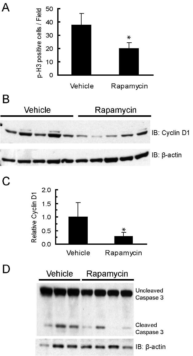
A. Rapamycin decreases the number of cells expressing phosphorylated histone-H3. The graph shows the average number of phosphorylated histone-H3 positive cells per field for primary tumors collected from vehicle (n=5) and rapamycin (n=6) treated mice. Five fields with equal numbers of cells/field were counted per tumor. Error bars represent standard deviations, * = p<0.05. B. Rapamycin decreases cyclin D1 levels in primary tumors. Whole cell lysates (100 μg) of primary MMTV-c-Neu tumors from vehicle and rapamycin treated mice were resolved by SDS-PAGE and transferred to PVDF membrane. Membranes were probed with an anti-cyclin D1 antibody. β-actin was used as a loading control. C. Bands were quantified by densitometry and the graph depicts mean cyclin D1 levels normalized to β-actin for vehicle and rapamycin treated mice. Error bars represent standard deviations, * = p<0.05. D. Western blot of uncleaved and cleaved caspase-3 levels in tumors collected from mice treated with vehicle or rapamycin for 5 days. β-actin serves as a loading control.