
Figure 6. Patterning of the ventral and distal optic vesicle occurs normally in Fzd5−/−embryos.
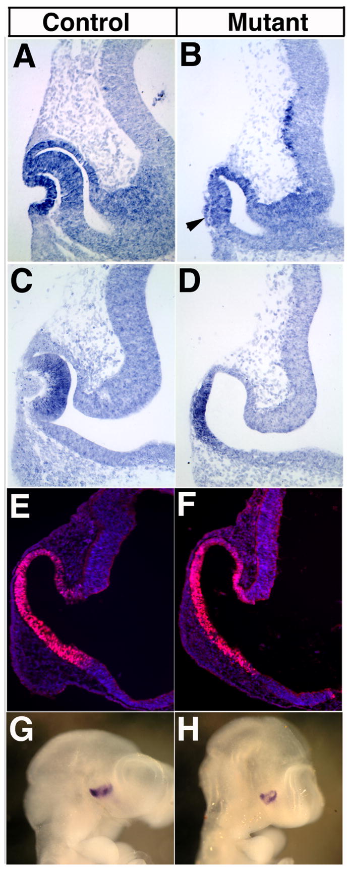
At E10.5, Pax6 is expressed in the developing neural retina, lens (arrowhead in B) and presumptive RPE of control (A) and Fzd5−/− embryos (B). Similarly, Chx10 is present in the neural retina of control (C) and in the distal region of the optic vesicle in mutant embryos (D). Pax2 protein is present in the optic vesicle and stalk at 27 somites in mutant embryos (E) similar to the expression observed in control embryos (F). Ventral expression of Vax2 is also normal in Fzd5−/− optic vesicles at 30 somites (compare G, H).