
Figure 4.
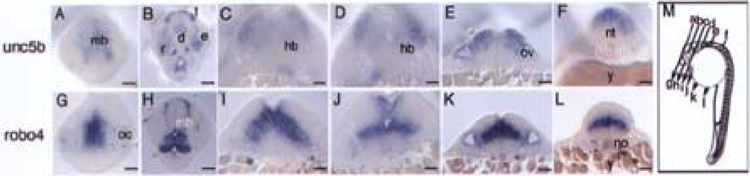
Comparative sectional analysis of embryonic brain regions of unc5b and robo4 in situ embryos. unc5b and robo4 24 hpf whole mount embryos were embedded in Spurr’s epoxy resin and sectioned as depicted in (M). All images were photographed at 40× magnification. Images were corrected for brightness, contrast, and sharpened in Adobe Photoshop. Scale bars: 50 μm. Abbreviations used include d: diencephalon, e: eye, hb: hindbrain, le: lens, mb: midbrain, no: notochord, nt: neural tube, oc: optic cup, ov: otic vesicle, r: retina, t: telencephalon, y: yolk.