
Figure 3.
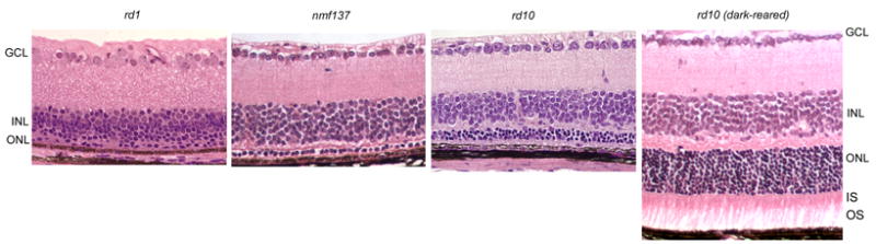
Histology of rd10, rd1, and nmf137 mouse retina at 24 days of age showed comparative degrees of retinal degeneration. By this stage, rd10 outer nuclear layer (ONL) of cyclic light reared mice was about four nuclei thick, whereas nmf137 was only one nuclei thick and rd1 had no photoreceptor nuclei. Conversely, ONL of dark-reared rd10 mice showed no thinning at 24 days of age and had substantial inner segments (IS) and outer segments (OS). Retinal ganglion cell layer (GCL) and inner nuclear layer (INL) did not vary with strain or lighting regimen.