
Figure 7.
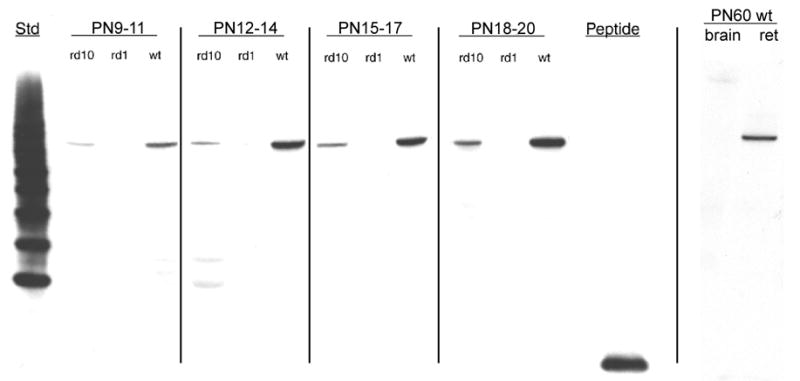
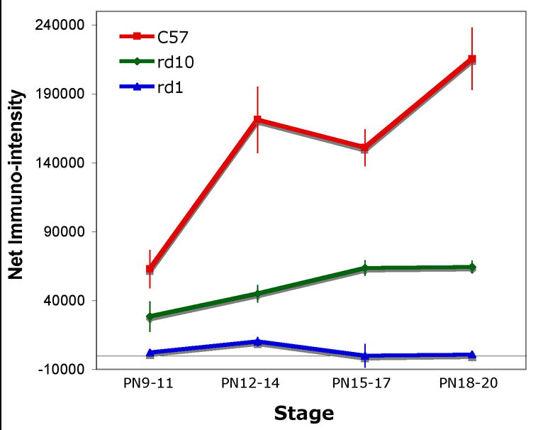
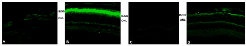
β-PDE immunoreactivity across development. Protein extracts from retinas removed from C57BL/6 (wild type; “wt”), rd1, and rd10 mice at postnatal days (P) 9 through 20 were analyzed by western immunoblotting using an antibody specific for the β subunit of rod cGMP PDE. Additionally, protein extract from retinas and whole brain of a P60 C57BL/6 were probed. Top Panel: Representative autoradiographs of western immunoblots. A band of approximately 86 kDa was present in the wt and rd10 retina protein extracts, but not in rd1 retina or wt brain extracts. Source for protein extracts is indicated above the lanes. “Std” is a “Magic Mark™” molecular weight standard (Invitrogen). “Peptide” is the synthetic peptide mix against which the primary antibody was raised (see text for details of this). As indicated by vertical lines, blots were taken from separate experiments and are representative. Middle Panel: Quantification of P9–20 data from western blots. Autoradiographs were scanned. Net intensities of bands were determined by subtracting background pixel densities from pixel densities of bands. Resulting net intensities were averaged for 3–4 mice per stage per strain. Bottom Panel: Immunohistochemistry shows specificity of β-PDE primary antibody. Cryosections of eyes from P22 rd1 (A), wt (B), and rd10 (D) mice were probed with the β-PDE primary antibody and FITC-conjugated secondary antibody. β-PDE antibody labeled the inner and outer segments of wt (B) and rd10 (D) photoreceptors, but no signal was apparent in rd1 sections (A) or in wt sections incubated with secondary antibody but not primary antibody (C).