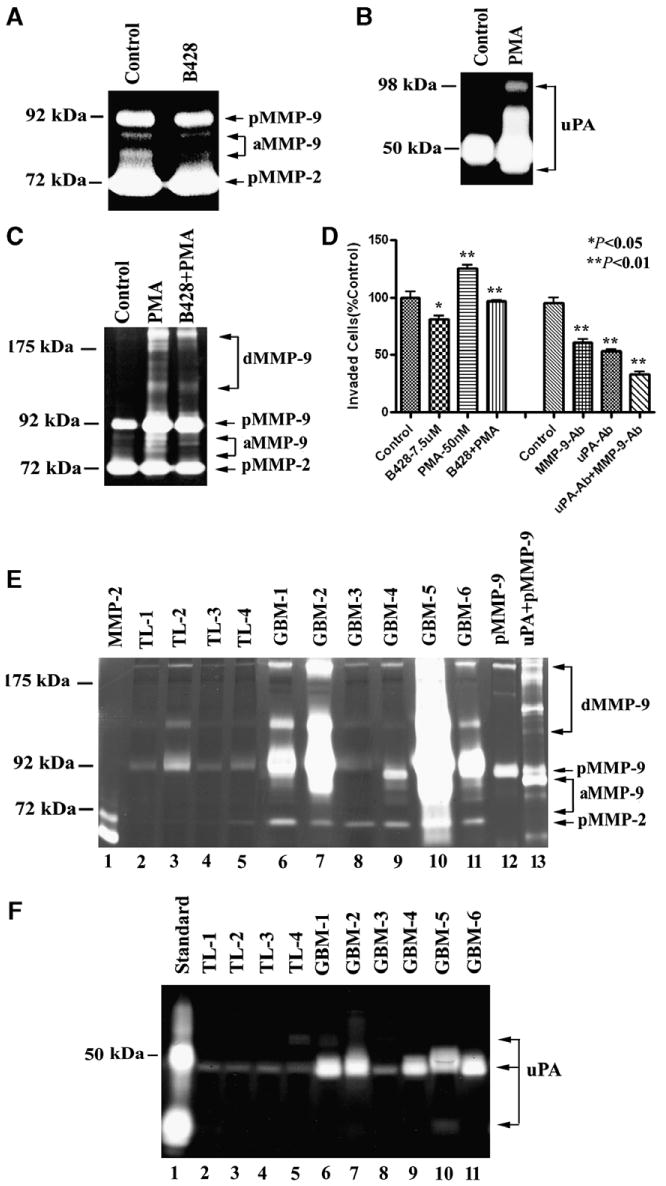
Fig. 4.

uPA-activated MMP-9 increased U1242 cell invasion and expression of uPA and MMP-9 in GBM patients' specimens and non-neoplastic temporal lobes (TL). (A) B428 (7.5 μM) reduced MMP-9 activity in U1242 cell media. pMMP-9, pro-MMP-9, aMMP-9, active MMP-9; pMMP-2. pro-MMP-2 (B) PMA (50 nM) induced uPA activity in U1242 cell media. (C) B428 (7.5 μM) blocked PMA-induced MMP-9 activity in U1242 cell media. dMMP-9, dimeric MMP-9 (D) Inhibition of uPA and MMP-9 activities reduced U1242 cell invasion. B428 (7.5 μM) blocked both U1242 and PMA-induced U1242 cell invasion. Combination of functional neutralizing MMP-9 and uPA antibodies more significantly inhibited U1242 cell invasion through fibronectin than uPA or MMP-9 antibody alone. The concentration of antibody was 25 μg/ml. Normal IgG was used as control. The numbers of control were used as the 100% invasiveness. Data shown are means ± SEM values from three separate experiments for each group. *p < 0.05; **p < 0.01. (E) Gelatin zymography analysis of MMP-2 and MMP-9 activities in TLs and GBMs. The recombinant MMP-2, latent MMP-9 and uPA-activated MMP-9 were used as controls. (F) Fibrinogen zymography analysis of uPA activities in TLs and GBMs. Recombinant uPA was loaded as control. Equal amount of protein (25 μg) was loaded each lane.
