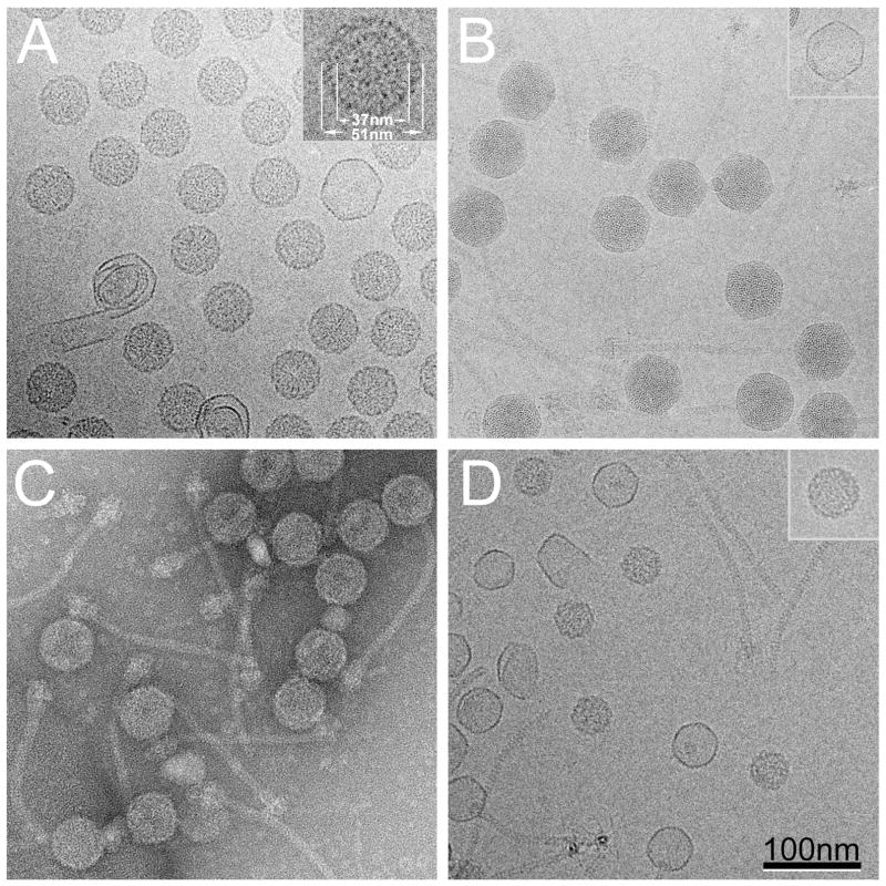
Figure 2.
(A) Cryo-EM of sucrose gradient–purified 80α procapsids. Inset, 2× magnified view of one procapsid with dimensions of the shell and the inner core indicated. (B) Cryo-EM of CsCl-purified 80α virions. The 2 nm spacing of the internal DNA is clearly visible. One thin-walled, empty capsid is also shown (inset). (C) Negatively stained CsCl-purified 80α procapsid fraction, containing a mixture of procapsids, tails and procapsids with attached tails. (D) Cryo-EM of sucrose gradient-purified SaPI1 procapsids. Inset, one of the about 5% 80α-size procapsids found in the SaPI1 procapsid sample. Scale bar, 100 nm.

