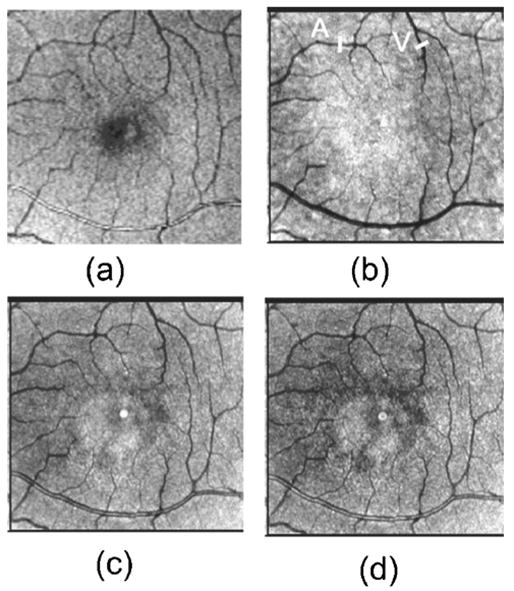
Fig. 4.

Retinal images of 66 year old Japanese man with a normal retina. (a) SLO image at 514 nm, (b) depolarized light images, (c) average reflectance image, (d) parallel polarized light image. SLO image at 514 nm, average reflectance image, and parallel polarized light image demonstrating the reflection at the center of the major retinal artery. This reflection was significantly reduced in the depolarized light images. Bar indicates the sample region for average vessel profiles of 11 bisector lines of retinal artery (A) and retinal vein (V). Bright spot in the center of each polarimetry image is an artifact due to internal reflections in the GDx-N.
