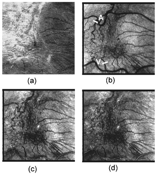
Fig. 6.

Retinal images of 70 year old Japanese man with an ERM. (a) SLO image at 514 nm, (b) depolarized light images, (c) average reflectance image, (d) parallel polarized light image. ERM could be clearly visualized as the highly reflected area in the SLO image at 514 nm. In the depolarized light image, the retinal vessels could be clearly visualized in the area of the ERM. In the average reflectance image and parallel polarized light image, the retinal vessels were somewhat obscured by the ERM. Bar indicates the sample region for average vessel profiles of 11 bisector lines of retinal artery (A) and retinal vein (V). Bright spot in the center of each polarimetry image is an artifact due to internal reflections in the GDx-N.
