Fig. 9.

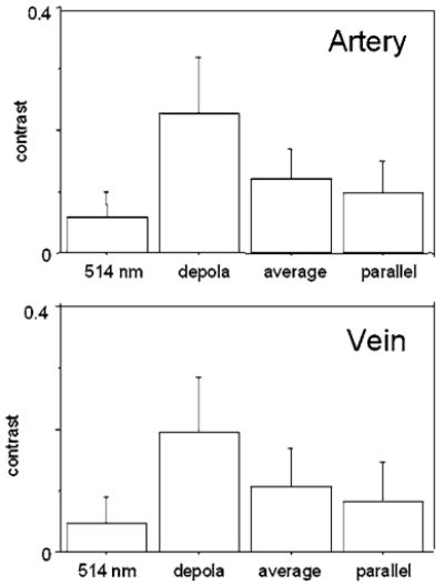
Michelson contrast of retinal arteries and veins in the eyes with ERMs. Contrast is greatest in the depolarized light image. Error bars indicate standard deviation of Michelson contrast.

Michelson contrast of retinal arteries and veins in the eyes with ERMs. Contrast is greatest in the depolarized light image. Error bars indicate standard deviation of Michelson contrast.