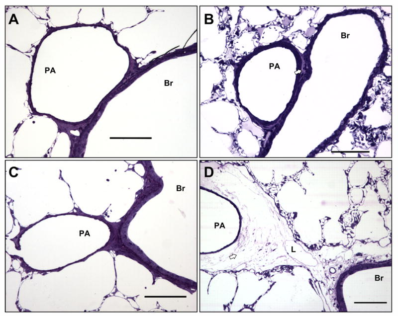
Figure 3.

Microscopic assessment of perivascular cuffing in extra-alveolar vessels. Perivascular cuffs were infrequently observed in control rat lungs (A). Perivascular cuffing induced by 4αPDD (B), 14,15-EET (C), or thapsigargin (D) was heterogeneous, and when cuffing appeared (arrow), cuff volume fraction was no different between groups (see Table 2). Extra-alveolar vessels often appeared no different control. Scale bars are 100 μm. PA, pulmonary arteriole; Br, bronchiole; L, lymphatic.
