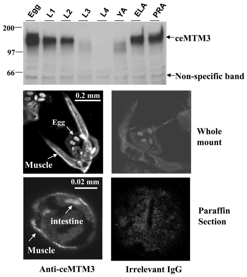
Fig. 1. Expression and distribution of ceMTM3 in C. elegans.

Top panel: C. elegans at different stages including egg, L1, L2, L3, L4, YA (young adult), egg-laying adult (ELA), and post-reproductive adult (PRA) were collected according to synchronized culture procedures. Cell extracts containing equal amounts of total proteins were subjected to Western blotting analysis with anti-ceMTM3 antibody. The position of ceMTM3 is indicated. A weak, non-specific band of ~60 kDa essentially serves as an internal loading control. Middle and bottom panels: whole mount (middle panel) and paraffin-embedded cross sections (bottom panel) of adult C. elegans were subjected to indirect immunofluorescent staining with anti-ceMTM3 antibody or irrelevant rabbit IgG. Arrows highlight positive staining at body wall muscle, eggs, and intestine.
