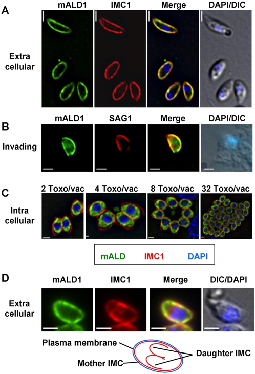
Figure 5. Distribution of aldolase-1 in extracellular, invading, and intracellular Toxoplasma.
(A–D) Immunofluorescence microscopy was used to determine the distribution of myc-tagged aldolase-1 (mALD1, green) in (A, D) extracellular parasites, (B) invading parasites, and (C) intracellular parasites immediately after invasion or after 1, 2, 3, and 5 rounds of replication. Panel D demonstrates the distribution of aldolase-1 in extracellular parasites caught in the process of endodyogeny. Note the selective association of the enzyme with only the IMC of the mother parasite and not that of the immature daughter cells. All panels show parasites and parasite-infected cells fixed in −20°C methanol. In panels A and D, parasites were counterstained with antibodies to the membrane skeleton protein IMC1 (red). In panel B, parasites expressing myc-aldolase-1 were allowed to interact with HFF cells for 2 minutes at 37°C, followed by the decoration of extracellular parts of the parasites with anti-SAG1 antiserum (red), which was in turn followed by cell fixation and permeabilization and staining with anti-myc monoclonal antibody (green). Parasite nuclei were visualized using DAPI (blue). Bars = 2 µm.

