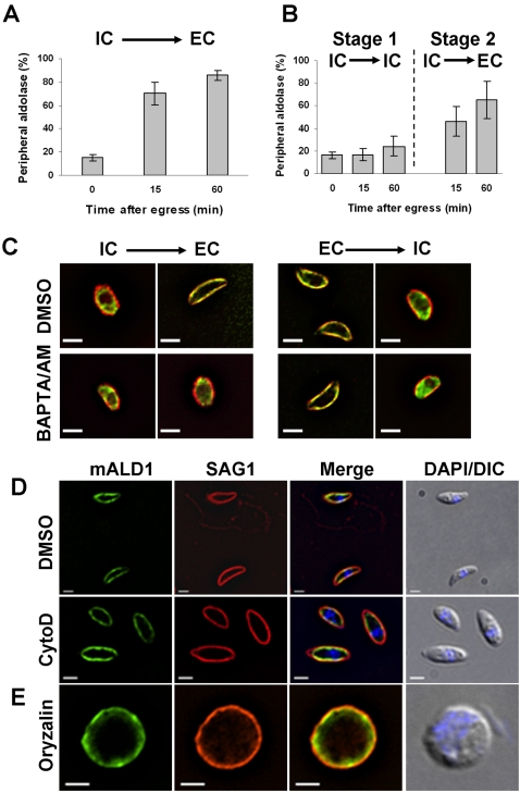
Figure 7. Aldolase-1 relocation during egress requires a decrease in environmental [K+] and an increase in [Ca2+]c and does not require either F-actin or microtubules.
(A) Intracellular parasites expressing myc-aldolase-1 were harvested 24 hours after infection in IC buffer as described in Materials and Methods and subsequently incubated for the indicated times in IC buffer at 37°C. After 60 minutes in IC buffer, parasites were recovered by centrifugation, resuspended in EC buffer, and incubated at 37°C for the indicated time. The fraction of parasites with peripheral aldolase-1 was determined as described above. (B) Intracellular parasites expressing myc-aldolase-1 were harvested 24 hours after infection in EC buffer as described in Materials and Methods and subsequently incubated for the indicated times in EC buffer at 37°C before fixation and processing for immunofluorescence microscopy as in Figure 4. The fraction of parasites with peripheral aldolase-1 was determined as in panel A. (C) Intracellular parasites expressing myc-tagged aldolase-1 were released from host cells in IC buffer and subsequently switched to EC buffer. Extracellular parasites expressing myc-tagged aldolase-1 were resuspended in EC buffer and further switched to IC buffer. Each buffer was supplemented by DMSO (control) or BAPTA-AM (20 µM). Parasites were incubated in each buffer for 30 min at 37°C and processed for immunofluorescence using mouse anti-myc antibody (green) and rabbit anti-IMC1 antiserum (red). Bars = 2 µm. (D) Intracellular parasites expressing myc-tagged aldolase-1 were treated with DMSO or 10 µM cytochalasin D (CytD) for 15 min at 37°C and subsequently released from host cells by passage through a 25G needle. Motility assays were performed in the presence of DMSO or cytochalasin D as described in Materials and Methods. Parasites were labeled using anti-myc (mALD1, green) and anti-SAG1 (red) antibodies. In the top panel, note the presence of a motile (top) and a non-motile (bottom) parasite. No motility was detected after cytochalasin D treatment. Overlay pictures of DIC and DAPI are shown on the right. Bars = 2 µm. (E) Intracellular parasites were treated for 24 hours with 2.5 µM oryzalin and subsequently released from host cells by passage through a 25G needle into EC buffer. After 15 minutes at 37°C, parasites were fixed and processed for immunofluorescence microscopy using anti-myc (mALD1, green) and anti-IMC1 (red) antiserum. An overlay of the DIC and DAPI are shown on the right. All samples were fixed in −20°C methanol. Bar = 2 µm.

