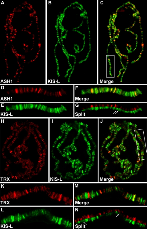
Figure 2. KIS-L co-localizes with the trithorax group proteins ASH1 and TRX on polytene chromosomes.
A–C) The distributions of ASH1 (A, red) and KIS-L (B, green) on wild-type polytene chromosomes are shown together with the merged image (C). D–G: Magnification of the chromosome arm bounded by the white rectangle in C is shown. Arrows in G mark examples of KIS-L bands that do not overlap with ASH1. H–J) The distributions of TRX (H, red), KIS-L (I, green) and the merged image (J) are shown. K–N: represent the magnification of the chromosome arm bound by the white rectangle in J. The arrows in N represent bands of KIS-L that do not overlap with TRX.

