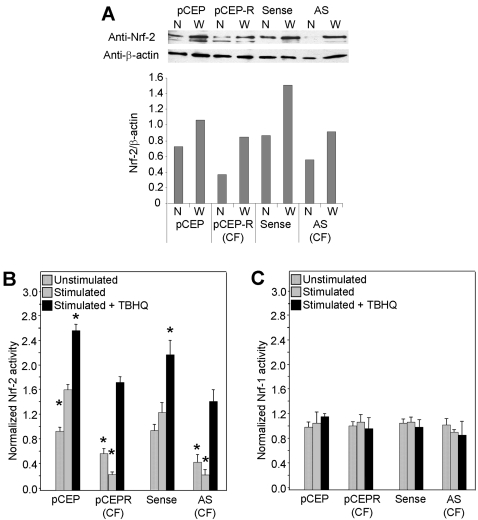
Figure 4. Nrf-2 expression and activity are decreased in CF epithelia in the absence and presence of inflammatory stimulation.
Normal and CF matched cell pairs are co-transfected with either a plasmid coding for Firefly luciferase expression driven by a Nrf-2 or Nrf-1 promoter and one coding for Renilla luciferase driven by the CMV promoter. Panel A: Western analysis of nuclear (N) or whole cell (W) Nrf-2 and an anti-β actin control. Band densities are used to calculate the relative abundance chart. Panel B: Cells transfected with the Nrf-2 promoter luciferase construct. Panel C: Cells transfected with the Nrf-1 promoter luciferase construct. Two days following transfection cell homogenates were assayed for luciferase activity by luminometer. Unstimulated cells are compared to cells stimulated with TNFα/IL-1β (10 ng/ml each) alone, or in the presence of an activator of Nrf-2 but not Nrf-1, tBHQ. * connotes significant difference (p<0.05) from respective normal control stimulated with TNFα/IL-1β (10 ng/ml each) alone. Each data bar represents the average of 8 replicate wells in 3 experiments for B and C.

