
Fig. 3.
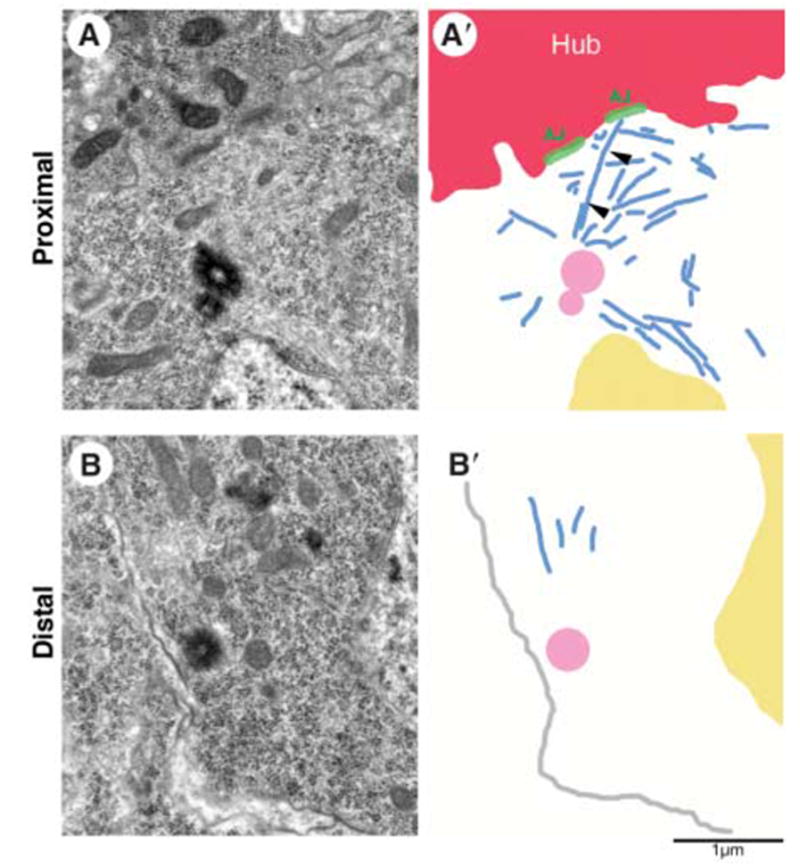
Centrosomes next to the hub harbor robust microtubule arrays. (A) Electron micrograph and (A′) summary diagram of a proximal centrosome in a GSC. Arrowheads in (A′) show a microtubule that runs from the centrosome to the adherens junction. (B) Electron micrograph and (B′) summary diagram of a distal centrosome in a GSC. Red, hub; blue, microtubules; green, adherens junctions; yellow, nucleus; pink, centriole; gray in (B′), plasma membrane. Scale bar, 1 μm.