Fig. 2.
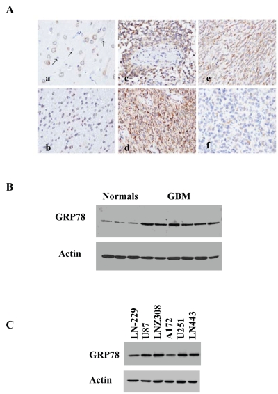
Expression of GRP78 in glioblastoma (GBM) specimens and glioma cell lines. (A) GRP78 expression in normal brain and GBM tissue sections: immunohistochemical staining of normal adult cortex with an anti-GRP78 antibody demonstrating cytoplasmic immunoreactivity of neurons and negative immunoreactivity of astrocytes (a); section of normal subcortical white matter showing positive GRP78 immunostaining of oligodendrocytes (b); anti-GRP78 immunohistochemical staining of human GBMs demonstrating a range of GRP78 immunoreactivity from strong diffuse positivity (c and d), to moderate diffuse positivity (e), to weak focal positivity (f). All original magnifications, ×400. The expression of GRP78 in normal brain and GBM specimens (B) and in glioma cell lines (C) was also examined using Western blot analysis.

