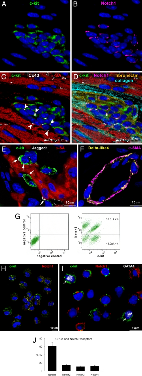
Fig. 1.
CPCs, c-kit, and Notch1 receptor. (A–D) Cardiac niche in which CPCs express c-kit (A, C, and D, green) and Notch1 (B and D, magenta). Connexin 43 (C and D, white dots, arrowheads) is present between CPCs and between CPCs and myocytes (C and D, α-sarcomeric actin, α-SA, red). The interstitium contains fibronectin (D, yellow) and collagen (D, light blue). (E) Jagged1 (white, arrows) is present between CPCs (c-kit, green) and myocytes. (F) Delta-like4 (yellow) is detected in the wall of a small coronary vessel (α-smooth muscle actin, α-SMA, magenta). (G) Dot plots showing the distribution of c-kit and Notch1 in CPCs. (H) Cytospin of FACS-sorted CPCs illustrating the coexpression of c-kit (green) and Notch1 (red). (I) CPCs (c-kit, green) that are positive for Notch1 (red) do not express GATA4. Two CPCs that are negative for Notch1 express GATA4 (white dots, arrows). (J) Distribution of Notch1–4 isoforms in CPCs.

