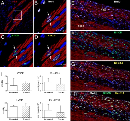
Fig. 5.
Myocardial regeneration. (A–D) The area included in the square (A) is illustrated at higher magnification in B–D. Regenerated myocytes (B–D: α-SA, red, arrows) are BrdU-positive (B, white), N1ICD-positive (C, green), and Nkx2.5-positive (D, yellow). (E–H) Area of regeneration (new) located within the infarct (dead). New myocytes are BrdU-positive (E), N1ICD-positive (F), and Nkx2.5-positive (G). (H) Merge of E–G. (I) Cardiac function in untreated and treated mice.

