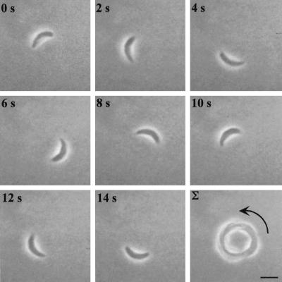
Figure 1.
Time-lapse video microscopy of a Toxoplasma cell engaged in circular gliding. Freshly harvested parasites in HHE medium were put on BSA-coated glass-bottom microwell plates, and their motility was documented with phase-contrast video microscopy using a 63× lens. The temperature was maintained at 37°C throughout the experiment using a temperature-controlled stage. The time elapsed between each frame is indicated in seconds. The final panel shows a frame-summation of the video, with the arrow indicating the net direction of movement. Bar, 6 μm. The video is shown at 2× real time and is representative of four independent experiments.

