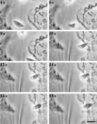
Figure 7.
Time-lapse video microscopy of a Toxoplasma tachyzoite lysing out from one host cell, move by helical gliding to a neighboring cell and invade. The time elapsed between each frame is indicated in seconds. The video was produced by capturing one frame per second and is shown at 2× real time. The invading parasite was centered by moving the microscope stage at the time of cell contact, which leads to a slight lag in the sequence. Because this sequence was filmed on an upright microscope, it is inverted with respect to those shown in the earlier figures. Bar, 6 μm.

