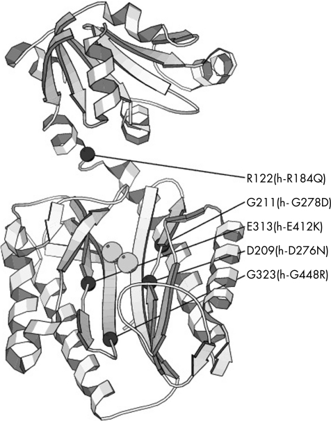
Figure 3 Position of the pathogenic mutations in the structure of Pyrococcus furiosus prolidase. The figure shows the ribbon representation of Pfprol (PDB entry 1PV9) together with the two metal ions bound to the active site (grey spheres). The positions of the C‐α atoms of the residues corresponding to the mutation sites in the human enzyme are shown as black spheres.

An official website of the United States government
Here's how you know
Official websites use .gov
A
.gov website belongs to an official
government organization in the United States.
Secure .gov websites use HTTPS
A lock (
) or https:// means you've safely
connected to the .gov website. Share sensitive
information only on official, secure websites.
