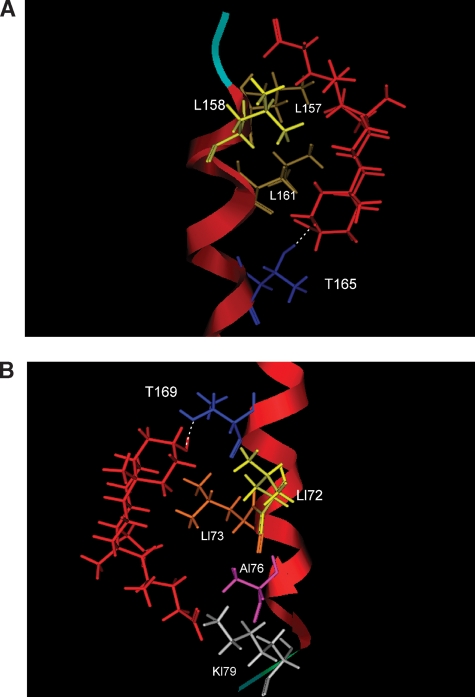
Fig. 6.
Putative models for LC docking on the BK β1 subunit transmembrane domain 2 (TM2). Molecular dynamics simulation (see Methods) indicates that LC interaction with one or two distinct regions in the channel β1 subunit TM2 may occur. In the “165/161 model” the ionized carboxylate in the side chain of LC resides in or near the extracellular aqueous compartment (A), whereas in the “169/172/173 model” the ionized carboxylate in the side chain of LC resides in or near the cytosolic aqueous compartment (B). The TM2 α helix is shown in red, whereas peptide backbone near the aqueous face is shown in pale blue (A) and green (B); selected β1 amino acids are shown in colors: T (dark blue), L (different types of yellow), A (purple), and K (light gray); LC is shown in red.

