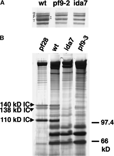
Figure 2.
SDS-PAGE analysis of dynein polypeptides in wild-type and mutant axonemes. (A) Shown here is the high molecular weight region of a 3–5% polyacrylamide gradient loaded with whole axonemes from wild-type, pf9–2, and ida7. The outer arm DHCs (α, β, and γ) are indicated on the left in wild type. The 1α and 1β DHCs of the I1 complex migrate between the β and γ DHCs of the outer arm, as indicated by asterisks on the right. Note that both pf9–2 and ida7 axonemes lack the 1α and 1β DHCs. (B) Sucrose density gradient centrifugation of dynein extracts from pf28, wild-type, ida7, and pf9–3. Shown here is a 5–15% polyacrylamide gradient gel loaded with fractions from the 19S region. This region typically contains the peak of the I1 complex and the trailing edge of the outer arm components that peak at 21S. The three ICs associated with the I1 complex are indicated on the left in the pf28 sample, which lacks the outer arms (Mitchell and Rosenbaum, 1985). The I1 ICs are also present in the wild-type sample, but are missing in both ida7 and pf9–3.

