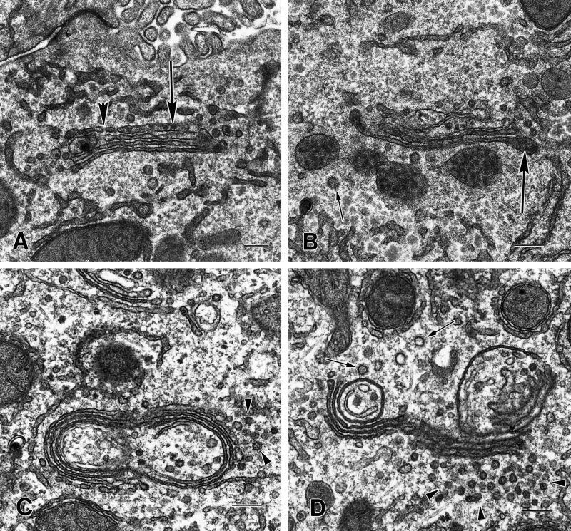
Figure 3.
Morphology of the compact regions of the Golgi ribbon in hepatocytes from CTL and CHX-treated animals. (A and B) The cisternae of the compact region of the Golgi ribbon from CTL animals are relatively straight. The cis cisternae (cis) were identified by their fenestrations (A, large arrowhead). Lipoprotein particles (large arrows) are present in the cisternae, particularly in the distended rims and/or vesicles in the trans region. Clathrin-coated vesicles (small arrow) are present in the trans region (B). (C and D) In the compact region of the Golgi stack the cisternae from CHX-treated animals are more tightly packed together and the width of the cisternal lumen is reduced when compared with CTLs. Lipoprotein particles are absent from the cisternae and vesicles. There are a large number of what appear to be vesicles (arrowheads), 50–70 nm in diameter, associated with the Golgi. Often the Golgi cisternae appear to be circular; this could result from a disruption at the noncompact region of the Golgi ribbon and some cisternae of the compact region folding back on themselves. The circularized Golgi cisternae are surrounded (both inside and out with many vesicles or tubules, some with clathrin-coats (small arrows, D). Bars, 200 nm.

