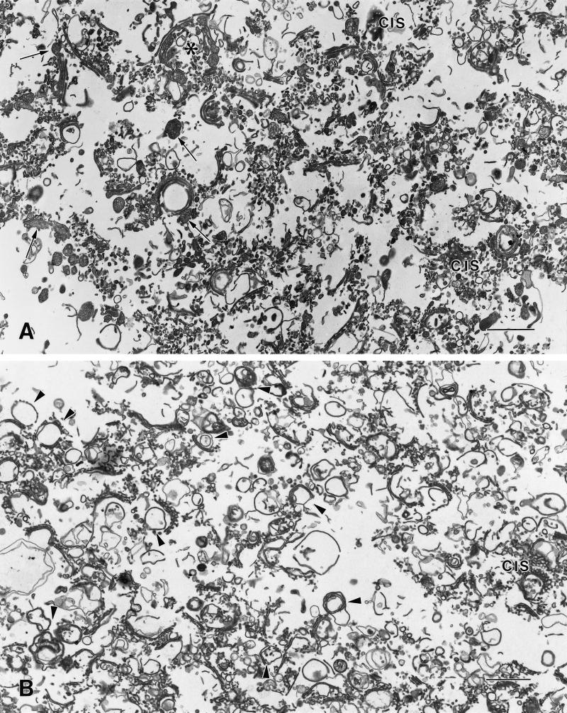
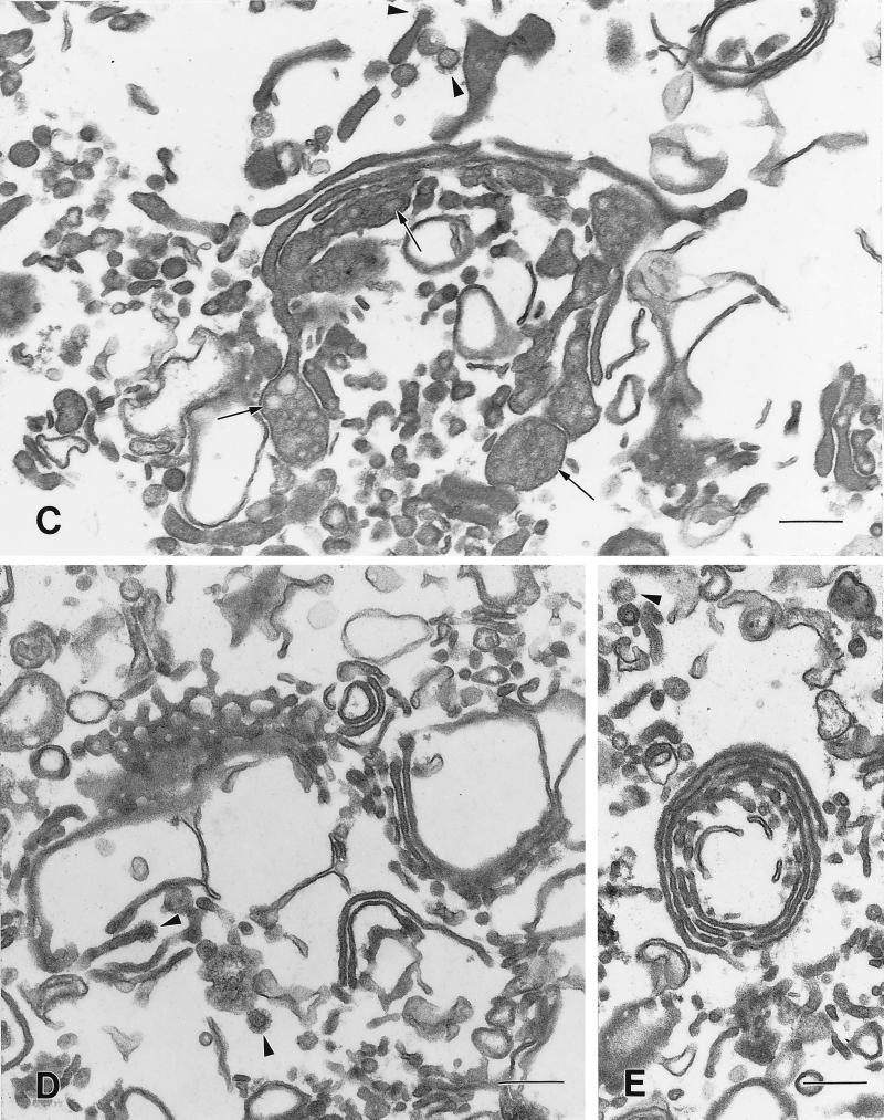
Figure 4.
Overview of SGF1s isolated from livers of CTL and CHX-treated animals. Low-magnification overview of CTL (A) and CHX (B) SGF1s. The majority of the components of the fractions are Golgi stacks, single cisternae, and associated vesicles. (A) In CTL SGF1, the cisternae are distended and contain secretory products (arrows). Most of the stacks are in a linear ribbon, although a few stacks appear circular. The asterisk indicates a Golgi region shown at higher magnification in C. (B) In CHX SGF1, the isolated stacks are condensed and do not contain obvious luminal secretory products. Compared with CTL SGF1, a higher proportion of the cisternae appear to be circular (arrowheads), similar to that observed in situ. The anastomosing tubular pattern of cis cisternae (cis) is particularly evident in the CHX SGF1. Higher magnifications of Golgi compact zones in CTL SGF1 (C) and CHX SGF1 (D and E). The stacked regions in the isolated CTL and CHX SGF1 have three or four cisternae. Arrowheads denote clathrin-coated structures. (C) In CTL SGF1, lipoprotein particles (arrows) are evident in the stacked cisternae, the dilated rims of the cisternae, and what appear to be adhering vesicles in the trans region. (D and E) In CHX SGF1, the cisternae of the Golgi stacks are more tightly packed and have reduced luminal widths when compared with the CTL. Lipoprotein particles are absent from the cisternae and vesicles. Circular profiles and cis regions are prominent. Bars: A and B, 1 μm; C–E, 200 nm.


