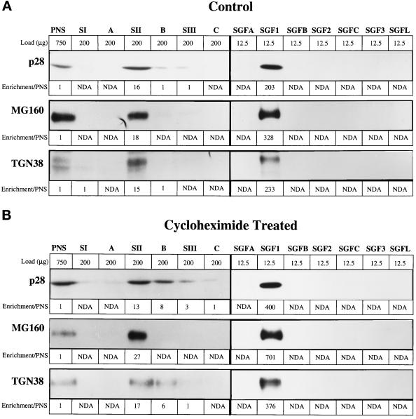
Figure 5.
Enrichment of Golgi markers in the stacked Golgi fraction. Golgi markers were analyzed in the fractions of the SGF1 isolation protocol by quantitative immunoblot of fractions from CTL (A) and CHX-treated (B) animals. The fractions are noted at the top and directly below is the load (micrograms of protein) of that fraction. It was necessary to increase the protein load of fraction in which the markers were present in low concentrations to obtain a reliable signal. For each marker, the values for enrichment over PNS are placed below the gel bands: cis-, p28; medial-, MG160; trans- and TGN, TGN38.

