
Figure 3. Atomic structure of SusD.
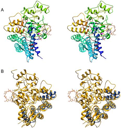
(A) Stereo ribbon diagram of apo-SusD, color-ramped from dark blue to red as the chain extends from the amino to the carboxyl end of the protein. An ordered Ca2+ ion is represented by a magenta-colored sphere while polyethylene glycol and ethylene glycol are shown as ball-and-stick figures. As a reference, a molecule of maltoheptaose from the structure of the SusD-maltoheptose complex is shown as a transparent ball-and-stick. (B) Stereo figure of SusD (yellow) highlighting residues 31-172 of PilF (blue) which contain the TPR units.