
Fig. 2.
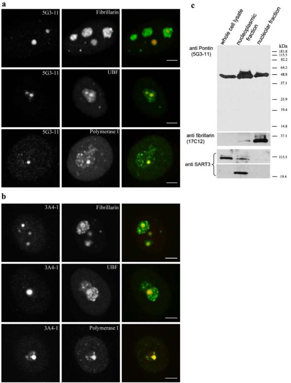
Pontin is localized in nucleoli. Double-labeling of HeLa cells with anti-Pontin antibodies 5G3-11 a or 3A4-1 b and anti-fibrillarin, anti-UBF or anti-RNA polymerase I antibodies. The large Pontin dots co-localize with UBF and RNA polymerase I containing dots. In contrast, Pontin revealed only partial co-localization with fibrillarin, which often formed a rim around Pontin dots. Note that in contrast to the three standard nucleolar proteins, Pontin dots appear only in a subset of nucleoli. Scale bar represents 5 μm. c Whole-cell lysate, nucleoplasmic and nucleolar fractions were prepared and used for immunoblotting with the anti-Pontin (5G3-11) antibody. Purity of the nucleolar fraction was verified by detection of a nucleolar marker fibrillarin and absence of the SART3 protein, which is present only in the nucleoplasm and devoid of the nucleolus. SART3 was partially cleaved during the nucleolar preparation and a lower molecular weight band appeared in the nucleoplasmic fraction