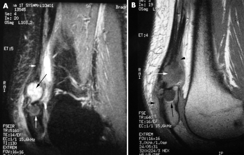
Figure 3 (A) Sagittal T2 weighted magnetic resonance image showing encysted fluid (black arrow) within the distal quadriceps tendon (arrowhead). It is closely applied to a large erosion in the superior pole of the patella (white arrow). (B) Sagittal T1 weighted magnetic resonance image showing an encysted area of intermediate signal (fluid) within the distal quadriceps tendon (white arrow). There is a large erosion (black arrow) within the superior pole of the patella. Oedema is present in the adjacent soft tissue (arrowheads). Patient consent has been obtained for publication of this figure.

An official website of the United States government
Here's how you know
Official websites use .gov
A
.gov website belongs to an official
government organization in the United States.
Secure .gov websites use HTTPS
A lock (
) or https:// means you've safely
connected to the .gov website. Share sensitive
information only on official, secure websites.
