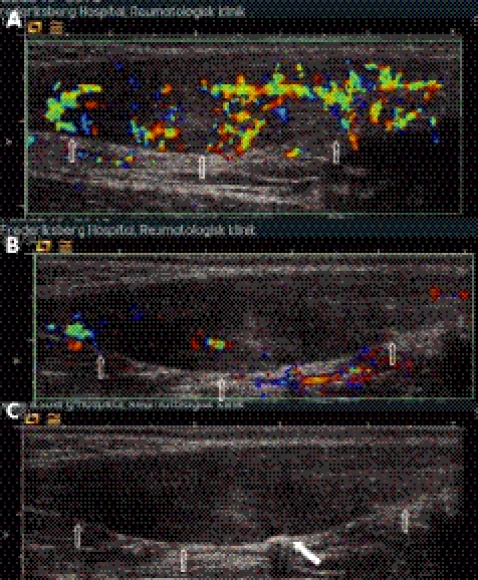
Figure 2 Colour Doppler activity at baseline and during treatment. Longitudinal images of an Achilles tendon with proximal oriented left. The anterior borders of the tendons are indicated with arrows. In (A) the Doppler activity was graded as 2 and had a colour fraction of 33%. In (B) and (C) the same tendon is being treated with coagulation therapy. The distal part of the tendon is “shut down” and the proximal part remains to be treated (B); (C) is the corresponding grey scale image of (B). The white filled arrow marks the ongoing coagulation.

An official website of the United States government
Here's how you know
Official websites use .gov
A
.gov website belongs to an official
government organization in the United States.
Secure .gov websites use HTTPS
A lock (
) or https:// means you've safely
connected to the .gov website. Share sensitive
information only on official, secure websites.
