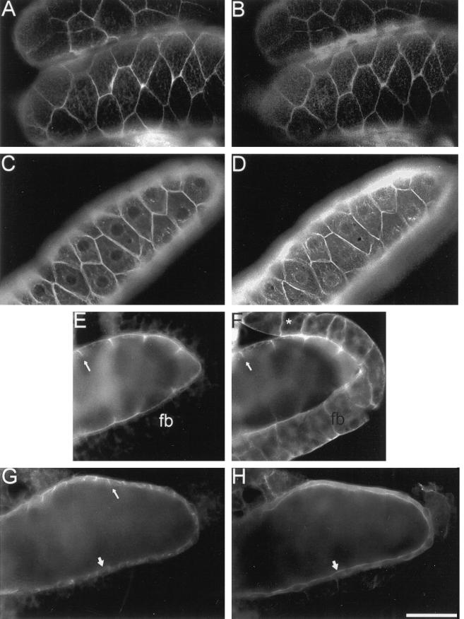
Figure 5.
Distribution of neuroglian, ankyrin, Na,K-ATPase, and βH spectrin in the larval salivary gland epithelium. Salivary glands were dissected from late third instar wild-type larvae, fixed, permeabilized and double labeled with mouse antineuroglian (A), rabbit anti-Drosophila ankyrin (B, D, and F), mouse anti-Na,K-ATPase (C, E, and G); and rabbit anti-Drosophila βH spectrin (H). Mouse antibodies were detected with Texas Red-conjugated secondary antibodies (A, C, E, and G) and rabbit antibodies were detected with FITC-conjugated secondary antibodies (B, D, F, and H). Neuroglian, ankyrin, and Na,K-ATPase colocalize at lateral sites of cell–cell contact (A–D). Ankyrin and the Na,K-ATPase colocalize at the lateral margin of salivary gland cells (arrow, E and F). Ankyrin is also concentrated at sites of cell adhesion (*, F) in the fat body (fb). Although the Na,K-ATPase was concentrated at lateral sites of cell contact in the salivary gland (thin arrow, G), the βH subunit of spectrin was concentrated at the apical cell surface (wide arrow, H). Bars: A–D and G–H, 100 μM; E and F, 67 μm.

