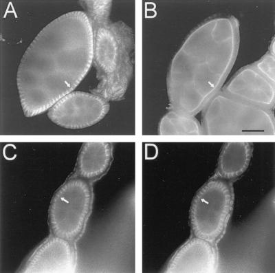
Figure 6.
Distribution of ankyrin (A and C), βH spectrin (B), and neuroglian (D) in the somatic follicle epithelium of the adult ovary. Ovaries were dissected from adult Drosophila females, fixed, permeabilized, and stained as in Figure 5. Arrows mark staining of ankyrin at lateral cell contacts (A and C), neuroglian at cell contacts (D), and βH spectrin at the apical surface (B). Bar, 20 μm.

