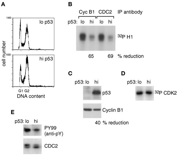
Figure 2.
Cell cycle distribution and analysis of CDC2/cyclin B1, p53, and CAK in TR9-7 cells 22 h after the removal of mimosine. Cells synchronized in S phase by treatment with mimosine for 48 h were incubated for 22 h in the presence (lo p53) or absence (hi p53) of tetracycline. (A) Cell cycle distribution. DNA content was determined by FACS analysis. (B) CDC2 activity. Histone H1 was phosphorylated in vitro by the use of [γ-32P]ATP, and immunoprecipitates (IPs) were prepared either with a monoclonal antibody specific for cyclin B1 (Cyc B1) or with a polyclonal antiserum specific for the C terminus of CDC2. The products were analyzed by SDS-PAGE and autoradiography. The amount of radioactive phosphate incorporated into histone H1 when p53 was induced was determined with a PhosphorImager and compared with the amount in cells without p53 induction (% reduction). (C) Expression of p53 and cyclin B1. Western analyses are shown. The levels of cyclin B1, determined as described in MATERIALS AND METHODS, were compared in the absence and presence of tetracycline (% reduction). (D) CAK activity. Activity was determined by phosphorylation of recombinant CDK2 by cyclin H immunoprecipitates in the presence of [γ-32P]ATP. (E) Expression and phosphorylation of CDC2. Tyrosine phosphorylation of CDC2 was determined after immunoprecipitation and Western transfer by the use of anti-phosphotyrosine (PY99). Total CDC2 was detected with anti-CDC2.

