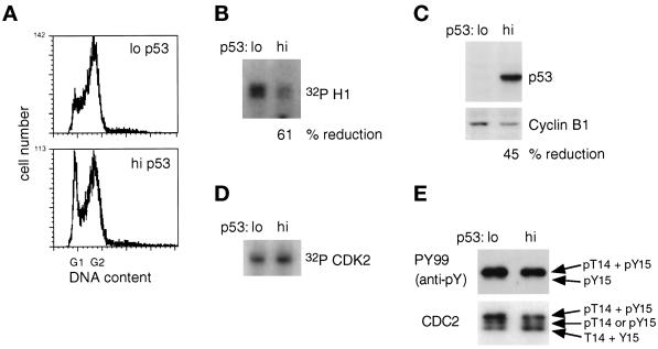
Figure 4.
Cell cycle distribution, cyclin B1 expression, and CDC2 activity in TR9-7 cells 28 h after the removal of mimosine in the presence of nocodazole. TR9-7 cells were released from a mimosine block in the presence (lo p53) or absence (hi p53) of tetracycline, and nocodazole was added 18 h later. Samples were analyzed after an additional 10 h. (A) Cell cycle distribution of TR9-7 cells after mimosine was removed and nocodazole was added. (B) CDC2 activity, assessed as described in Figure 3. (C) Expression of p53 and cyclin B1. The proteins were detected by Western transfer, and % reduction is shown. (D) CAK activity, assessed as described in Figure 2. (E) Tyrosine phosphorylation of CDC2 in TR9-7 cells 28 h after mimosine was removed. Phosphorylation of CDC2 on tyrosine was determined as described in Figure 2. pT, phosphothreonine; pY, phosphotyrosine.

