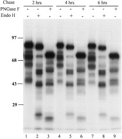
Figure 3.
Endo H and PNGase F digestion of HA fragments generated from addition of puromycin. Influenza-infected CHO cells were labeled as in Figure 1 in presence of 30 μM puromycin, and then chased for 2, 4, and 6 h in the presence of protease inhibitors. After the chase, cell lysates were collected and immunoprecipitated with HA antibodies. The precipitated proteins were digested with PNGase F at 37°C for 1 h to remove oligosaccharides or Endo H at 37°C overnight. The top band of each lane represents the full-length HA.

