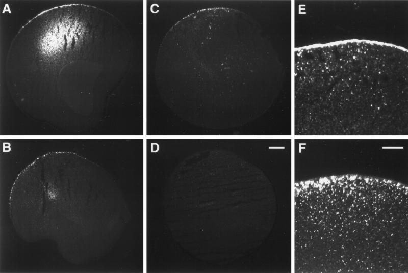
Figure 5.
Immunofluorescence detection of XENaCF with anti-FLAG antibody. (A) XENaCF-expressing control oocytes showed a bright signal over the plasma membrane of the vegetal pole and in an intracellular compartment. XK-Ras2AG12V–coinjected oocytes (B) as well as progesterone-treated control oocytes (C) showed a similarly polarized staining in the low-power magnification but in a dotted pattern. No signal was detected in uninjected oocytes (D). The high-power magnification revealed in XENaCF–injected oocytes a continuous staining over the plasma membrane and of a few submembranous dotted structures (E). In XK-Ras2AG12V–coinjected oocytes, a dotted staining pattern was localized almost entirely below the plasma membrane (F). Bars: D, 100 μm; F, 40 μm.

