
Fig.2. 4-HNE and anti-Fas CH11 antibodies-induced apoptosis in WT and CRL2571 Jurkat cells.
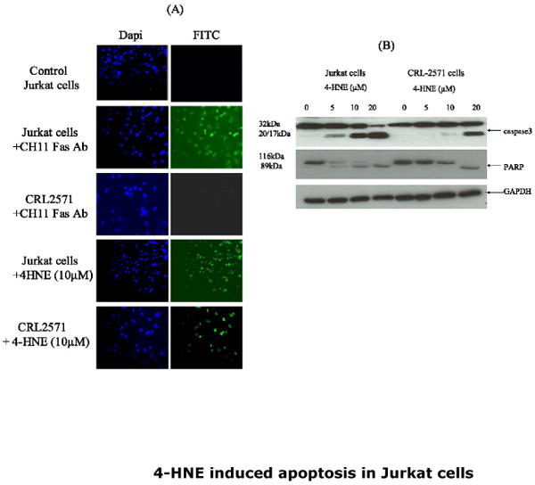
(A) In-situ activation of caspase3: Cells (2×10⁵) were either treated with anti-Fas CH 11 antibodies (250ng/ml) or with 10μM 4-HNE for 2h at 37°C in 8ml complete growth medium. The activation of caspase was examined by staining with 10 μM CaspACE FITC-VAD-FMK in situ marker as per the manufacturer's instructions. The slides were mounted with Vectashield DAPI mounting medium and observed under fluorescence microscope (Nikon). The photographs were taken with 20× objective lens. DAPI and FITC stained cells are appropriately marked in panels (B) Western blot analysis showing the activation of caspase3 and PARP cleavage: WT and CRL2571 Jurkat cells (2×10⁵) were separately treated with different concentrations of 4-HNE (0-20μM) for 2h at 37°C in complete growth medium. The cells were pelleted and washed 2× with PBS and the cell lysates were prepared as described in Materials and Methods section. Cell extracts (50μg protein) were then subjected to Western blot analysis using anti-caspase3 or anti-PARP antibodies. Anti-GAPDH antibodies were used as loading control.