
Fig.3. Effect of 4-HNE on the expression of Fas in CRL2571 cells.
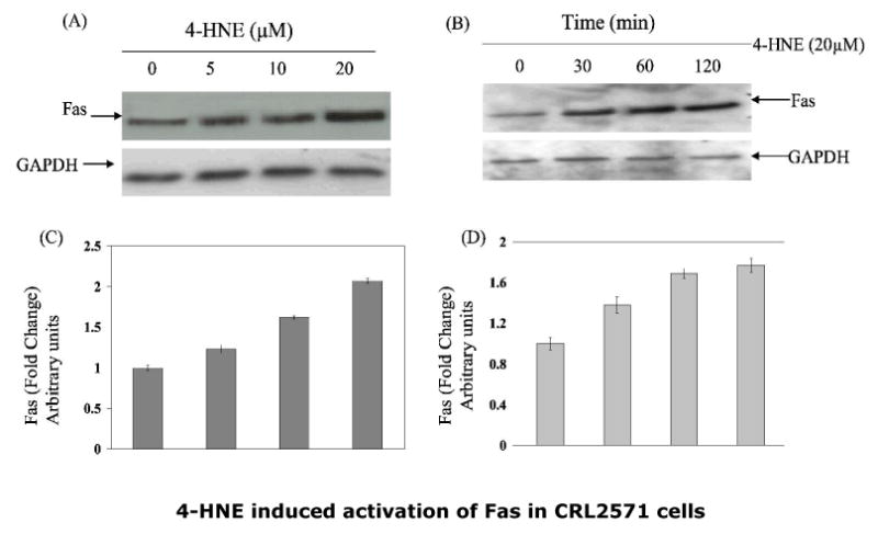
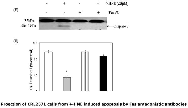
To examine the dose (panel A) or time-dependent (panel B) effect of 4-HNE, cells (2×10⁵) were treated with 4-HNE (0-20μM) for 2h or with 20μM 4-HNE at different time points (0-120min) in complete growth medium at 37°C. After incubation the cells were pelleted, washed 2× with PBS and homogenized in radioimmunoprecipitation assay (RIPA) buffer and centrifuged at 10,000g. Supernatants containing 50μg protein were subjected to Western blot analysis using anti-Fas monoclonal antibodies (B-10) as described in the Methods section. Blots were developed by West Pico-chemiluminescence reagent (Pierce). (C and D) Bar graphs showing the fold change (mean ± SD, n=3) in densitometric analysis of the Fas bands relative to GAPDH bands using Kodak 1D 3.6 image analysis software. (E) Effect of antagonistic anti-Fas antibodies on 4-HNE- induced caspase3 activation: Cells (2×10⁵) were plated in Petri-dishes and pretreated with anti-Fas (B-10) monoclonal antibodies (2μg IgG protein) for 2h followed by 20μM 4-HNE treatment for another 2h at 37°C. Cells were washed with PBS (2×) and counted in a hemocytometer using Trypan blue dye exclusion method. Rest of the cell pellets were extracted in RIPA buffer as described above and the extracts were analyzed for the activation of caspase3 on immunoblots using anti-caspase3 antibodies. (F) Attenuation of 4-HNE toxicity in anti-Fas antibodies (B-10) coated cells: Bar chart showing the percent cell survival in control and Fas antibodies treated cells after treatment with 4-HNE. Data presented are mean ± SD of two separate experiments done in triplicate. (* significantly different from the control cells p<0.01)